
Cell, mouse models in Angelman shed light on mechanism of disease
Increases seen in pathway for clearing cellular waste in study
Written by |

Increases in autophagy, a pathway for clearing cellular waste, were observed in cell and mouse models of Angelman syndrome (AS) in a recent study that revealed new details of this disease mechanism.
Researchers noted that it isn’t yet clear whether this increase is a compensatory process to help restore more normal brain function, or if it’s part of the disease-driving processes.
“Our results … may have broad implications,” not only for Angelman but for various diseases in which autophagy and other related mechanisms play a role, the scientists wrote.
The study, “UBE3A deficiency-induced autophagy is associated with activation of AMPK-ULK1 and p53 pathways,” was published in the journal Experimental Neurology.

More research needed to understand mechanism of disease
Angelman syndrome is a complex neurological disorder caused by a missing or dysfunctional copy of the UBE3A gene that people normally inherit from their mothers.
UBE3 is responsible for producing the UBE3A protein, a key player in the ubiquitin-proteasome system, called UPS, which degrades unneeded or damaged proteins.
Another way of getting rid of cellular waste is through autophagy, wherein waste is transported into cell compartments called lysosomes for recycling. Because autophagy is critical for normal cellular function, impairments in this process have been linked to a range of diseases. However, its role in Angelman hasn’t been established.
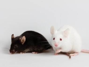
Now, researchers investigated whether autophagy is disrupted in mouse and cell models of AS.
Markers of increased autophagy were observed in the cell model relative to normal cells. They also were elevated in the cerebellum of AS mice compared with healthy mice. The cerebellum is one of the brain regions that’s mainly impacted in AS.
Importantly, evidence suggested that both the initiation of autophagy as well as its degradative activity were elevated in the AS samples.
“Together, these results indicate that UBE3A deficiency increases autophagy activity,” the researchers wrote.
The scientists then examined the levels of a number of other proteins to determine the mechanisms by which this autophagy boost may happen in AS.
A particularly notable finding was an elevation in an active form AMPK in AS mice relative to healthy mice.
While AMPK has a number of downstream targets and serves various cellular functions, it’s involved in the initiation of autophagy in part through its activation of the ULK1 protein.
Levels of active ULK1 were similarly elevated in AS mice, overall suggesting that the activation of AMPK and ULK1 may be central to the observed increases in autophagy, according to the researchers.
Another protein called p53, which both positively and negatively regulates autophagy, was found to have a non-significant increase in its levels in the nucleus, or core, of cells from AS mice. Meanwhile, its levels were significantly decreased in the cytoplasm, or the fluid surrounding the nucleus.
Those findings are consistent with p53’s known role of promoting autophagy in the nucleus and its ability to suppress autophagy when in the cytoplasm, the researchers noted. Again, these results favor conditions for autophagy, they noted.
“Further studies are needed to investigate whether [p53] depletion and inhibition could affect autophagy in both cell lines and in AS mice,” the researchers wrote.
Other observed changes could possibly contribute to the enhancement in autophagy seen, for example, in the movement of a molecule called TFEB into the cell’s nucleus.
Still, the researchers believe that the autophagy increase is “most likely caused by increased AMPK activation followed by ULK1 activation,” with possible contributions from other pathways.
Whether enhanced autophagy is a compensatory mechanism to sustain metabolic homeostasis or a pathogenic [disease-driving] process for Angelman syndrome remain to be determined.
Dysfunction of the UPS, as is seen in AS, has been previously linked to increases in autophagy. When the UPS can’t clear damaged proteins the way it usually does, it’s possible the body recruits the autophagy pathway to take on that role.
“Recent studies have revealed dynamic interconnections and metabolic compensation between these two systems,” the scientists wrote.
Still, “whether enhanced autophagy is a compensatory mechanism to sustain metabolic homeostasis or a pathogenic [disease-driving] process for Angelman syndrome remain to be determined,” the team concluded.

Leave a comment
Fill in the required fields to post. Your email address will not be published.