
Seizures, altered brain structures linked in Angelman children: Study
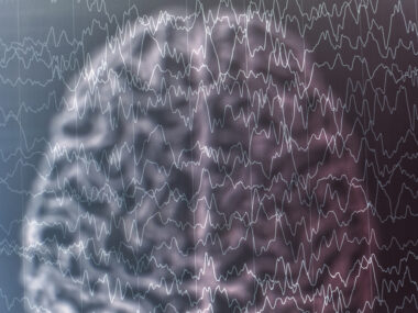
MRI brain imaging saw differences in the outer cortex layer, inner subcortex gray matter
Written by |

Certain structures within the brains of children with Angelman syndrome were altered compared with unaffected children of the same age, an MRI study concluded.
Imaging found a thinner and more folded outer cortex layer and reduced gray matter volume within the inner subcortex region in Angelman children.
More serious abnormal brain patterns appeared to be linked to the occurrence of seizures, the researchers noted in the MRI study, “Cortical and subcortical morphological alteration in Angelman syndrome,” which was published in the Journal of Neurodevelopmental Disorders.
Angelman syndrome is a neurodevelopmental disorder marked by poor muscle control (ataxia), intellectual disability, speech impairment, seizures, and hyperactivity.
Emerging evidence from MRI imaging studies indicates changes in several structures within the brains of Angelman patients, which may affect brain function and cause symptoms.
Researchers in China applied a set of high-resolution brain MRI measurements to detail the Angelman brain and determine whether any detected abnormalities are associated with seizures.
“To the best of our knowledge, this is the first study conducted on a Chinese population with [Angelman syndrome],” they wrote.

Brain differences and connection to seizure activity
The brain structures of 32 Angelman patients (16 girls, 16 boys), ages 4-9 and 26 age- and sex-matched healthy controls were compared by MRI. All the patients in this group had experienced seizures.
The researchers focused on the cortical region, the outer folded layer of the brain involved in higher functions, such as decision-making and language, as well as the subcortical region, the inner part with more primitive functions — emotional processing, memory, emotion, and pleasure.
Gray matter volume, cortical thickness, and the local gyrification index, which reflects the extent of cortical folding, were measured. The gray matter mainly contains nerve cell bodies.
Compared with controls, children with Angelman showed a significant decrease in gray matter volume in the cortical, subcortical regions, and the cerebellum at the back and base of the brain. In the brain area of the cortex called the inferior precuneus, the gray matter volume of Angelman patients was significantly increased, however.
Patients’ cortical region also showed more extensive folding across the whole brain than controls, and in some areas the cortex was thinner.
Researchers noted that more extensive folding of the brain’s cortex and increased gray matter volume in the inferior precuneus were “important findings.”
To investigate relationships between brain structures and seizures, a second group of 24 Angelman patients with seizures, ages 1-3, were compared with 17 age-matched patients without seizures.
The gray matter volume was reduced in both cortical and subcortical regions of patients with seizures compared with those without seizures. The cortical thickness was also reduced in seizure patients and there was a small increase in folded cortical structures in the right precuneus.
Researchers then focused on two areas significantly affected in Angelman patients — the precuneus in the cortex and the caudate in the subcortex. Each segment of the caudate, called the head, body, and tail, showed sections of lower density in seizure versus non-seizure patients.
The extent of cortical folding was significantly greater in the precuneus on both sides of the brain in patients compared with controls, while the cortical thickness was reduced in the right precuneus, but not the left. In comparison, the left precuneus showed a significant difference in thickness between the seizure and non-seizure groups.
“These results revealed cortical and subcortical morphological alterations in patients with [Angelman syndrome],” the researchers concluded. “The abnormal brain pattern was more serious in patients with seizures, suggesting that the occurrence of seizures may be related to abnormal brain changes.”


Leave a comment
Fill in the required fields to post. Your email address will not be published.